ABDOMINAL ULTRASOUND


-
An abdominal ultrasound image is a useful way of examining internal organs, including the liver, gallbladder, spleen, pancreas, kidneys, and bladder. Because US images are captured in real time, they can show movement of internal tissues and organs and enable physicians to see blood flow. This can help to diagnose a variety of conditions and to assess damage caused by illness.Ultrasound imaging is used extensively for evaluating the kidneys, liver, gallbladder, pancreas, spleen, and blood vessels of the abdomen. Because it provides real-time images, it can also be used to:
- Guide procedures such as needle biopsies, in which needles are used to sample cells from organs for laboratory testing.
- Help a physician determine the source of many abdominal pains, such as stones in the gall bladder or kidney, or an inflamed appendix.
- Help identify the cause for enlargement of an abdominal organ.
Doppler
ultrasound is a special type of ultrasound study that examines major
blood vessels. These images can help the physician to see and evaluate:
- Blockages to blood flow, such as clots.
- Build-up of plaque inside the vessel.
- Congenital malformation.
- With knowledge about the speed and volume of blood flow gained from an ultrasound image, the physician can often determine whether a patient is a good candidate for a procedure like angioplasty.
LIVER
Thirty
per cent of the blood pumped through the heart in one minute passes
through the body's chemical factory, which is called the liver. The
liver cleanses the blood and processes nutritional molecules, which are
distributed to the tissues. The liver also receives bright red blood
from the lungs, filled with vital oxygen to be delivered to the heart.
The only part of the body which receives more blood than the liver is
the brain. The liver is located at the top of the abdomen, just below
the diaphragm and has two main lobes. It is the largest gland in the
body, weighing 2.5 to 3.3 pounds. When we eat, more blood is diverted to
the intestines to deal with digestive processes; when not eating,
three-fourths of the blood supply to the liver comes from the intestines.
PANCREAS
The
pancreas is an elongated, tapered organ located across the back of the
abdomen, behind the stomach. The right side of the organ (called the
head) is the widest part of the organ and lies in the curve of the
duodenum (the first section of the small intestine). The tapered left
side extends slightly upward (called the body of the pancreas) and ends
near the spleen (called the tail). The pancreas is a gland organ in the
digestive and endocrine system of vertebrates. It is both an endocrine gland producing
several important hormones, including insulin, glucagon, and
somatostatin, and a digestive organ, secreting pancreatic juice
containing digestive enzymes that assist the absorption of nutrients and
the digestion in the small intestine. These enzymes help to further
break down the carbohydrates, proteins, and lipids in the chyme.
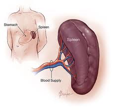
SPLEEN
The
spleen is the largest of the lymphoid tissues. It is just about the
size of the heart and is a spongy material which will hold up to .3
gallons of blood.This is an organ in the upper far left part of the
abdomen, to the left of the stomach. The spleen varies in size and shape
between people, but it’s commonly fist-shaped, purple, and about 4
inches long. Because the spleen is protected by the rib cage, you can’t
easily feel it unless it’s abnormally enlarged.
The spleen plays multiple supporting roles in the body. It acts as a filter for blood as part of the immune system. Old red blood cells are recycled in the spleen, and platelets and white blood cells are stored there. The spleen also helps fight certain kinds of bacteria that cause pneumonia and meningitis.
The spleen plays multiple supporting roles in the body. It acts as a filter for blood as part of the immune system. Old red blood cells are recycled in the spleen, and platelets and white blood cells are stored there. The spleen also helps fight certain kinds of bacteria that cause pneumonia and meningitis.

GALL BLADDER
Gall bladderis a small organ that aids mainly in fat digestion and concentrates bile produced by the liver. In humans, the loss of the gall bladder is usually easily tolerated. The surgical removal of the gall bladder is called a cholecystectomy.The gallbladder is a hollow system that sits just beneath the liver
In adults, the gall bladder measures approximately 8 centimetres
(3.1 in) in length and 4 centimetres (1.6 in) in diameter when fully
distended It is divided into three sections: fundus, body and neck.
Function
When food containing fat enters the digestive tract, it stimulates the secretion of cholecystokinin (CCK). In response to CCK, the adult human gall bladder, which stores about 50 millilitres (1.7 U.S. fl oz; 1.8 imp fl oz) of bile, releases its contents into the duodenum. The bile, originally produced in the liver, emulsifies fats in partly digested food.


KIDNEYS
About
one-quarter (750-1,000 pints daily) of the blood which is output by the
heart is sent to the body's "filter treatment plant", where it is
purified by the kidneys and circulated on to the rest of the body. One
to two thousandths (1/1000-2/1000) of the blood flow becomes fluid waste
and is sent into the bladder for storage until it can be conveniently
expelled. This toxic waste is called urine. The kidneys are located
about two inches above the body's midline just below and behind the
liver in the upper abdomen and behind the lower ribs. They receive about
120 pints of blood per hour, even if other body systems are shorted.
They are the balancers of internal fluids, so if we overeat or overdrink
one day and diet the next, or if we have an active, "sweaty" day, the
kidneys will compensate and see that these fluctuations in fluid, salt
and glucose are leveled out. It is important to drink plenty of fluids
each day to keep the kidneys in good working order. The "kidney" bean is
so named because of its resemblance to the shape and color of the
kidneys. Many people believe the kidneys lie down in the flanks and are
surprised when pain from kidney disorder comes from the upper middle
back area. Well, I have "to go" now.


No comments:
Post a Comment