A transducer converts one type of energy into another. Based upon the pulse-echo principle occurring with ultrasound piezoelectric crystals, ultrasound transducers convert:
– Electricity into sound = pulse
– Sound into electricity = echo
– Electricity into sound = pulse
– Sound into electricity = echo
• Pulse of sound is sent to soft tissues
• Pulsing is determined by the transducer or probe crystal(s) and is not operator controlled.
• Pulsing is determined by the transducer or probe crystal(s) and is not operator controlled.
• Echo produced by soft tissues
• Tissue interaction with sound =acoustic propagation properties
• Echoes are received by the transducer crystals
• Echoes are interpreted and processed by the ultrasound machine
• Tissue interaction with sound =acoustic propagation properties
• Echoes are received by the transducer crystals
• Echoes are interpreted and processed by the ultrasound machine
Transducer Types
• Mechanical
• Electronic
– Linear Arrays
– Curved Arrays
• Mechanical
• Electronic
– Linear Arrays
– Curved Arrays
– Phased Arrays
Linear - the linear array scanners produce sound waves parallel to
each other and produces a rectangular image. The width of the image and number
of scan lines are the same at all tissue levels. This has the advantage of good
near field resolution. Often used with high frequencies ie 7MHz. Can be used
for viewing surface texture of the liver. There disadvantage is artifacts when
applied to a curved part of the body creating air gaps between skin and
transducer.
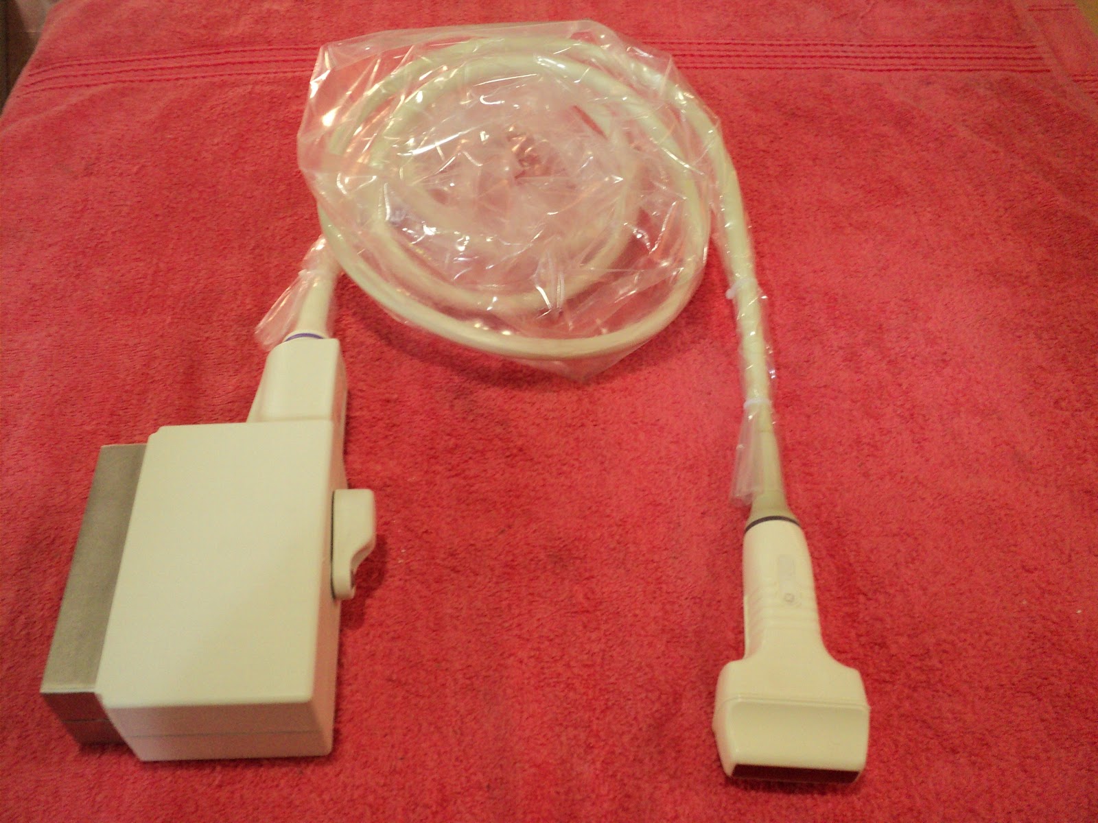 |
| 12L LINEAR PROBE Taken by, Ms.Praseetha |
 |
| 12L LINEAR PROBE Photo Taken by, Ms.Praseetha |
Sector/Vector - Produces a fan like image that is narrow near the
transducer and increase in width with deeper penetration. It is useful when
scanning between the ribs as it fits in the intercostal space. The disadvantage
is poor near field resolution.

 |
| 3S sector probe |
Curved - Often with frequencies of 2 - 5 MHz (to allow for a range
of patients from obese to slender). It is a compromise of the Linear and Sector
scanners. The density of the scan lines decreases with increasing distance from
the transducer. Can be difficult to use in curved regions of the body eg. the
spleen behind the left costal margin.
 |
 |
| taken by, C60e/5-2MHz Ms.Praseetha |
3D Transducers
Matrix Transducer

- 3 to 1 MHz extended operating frequency range
- 2D Matrix phased array with 2,400 elements
- 2D, biplane (Live xPlane), triggered full volume, Live 3D Echo, Color Doppler with 2D, biplane and 3D,Harmonic Imaging
Mechanical
3D Tranducer
- 6 to 2 MHz extended operating frequency range
- Supports high resolution 2D imaging
- high resolution, quantitative, single sweep 3D volume aquisition
- 4D imaging up to 36 volumes per second
- Color Doppler
- Field of view: 66 degrees
- General purpose abdominal, obstetrical and gynecological applications
 |
| RAB 2-5 PROBE |

No comments:
Post a Comment